ABOUT US
GET A FREE QUOTE
-
Home

-
Products
-
Ophthalmic Equipment
-
OCT
- OCT-8200 Optical Coherence Tomography
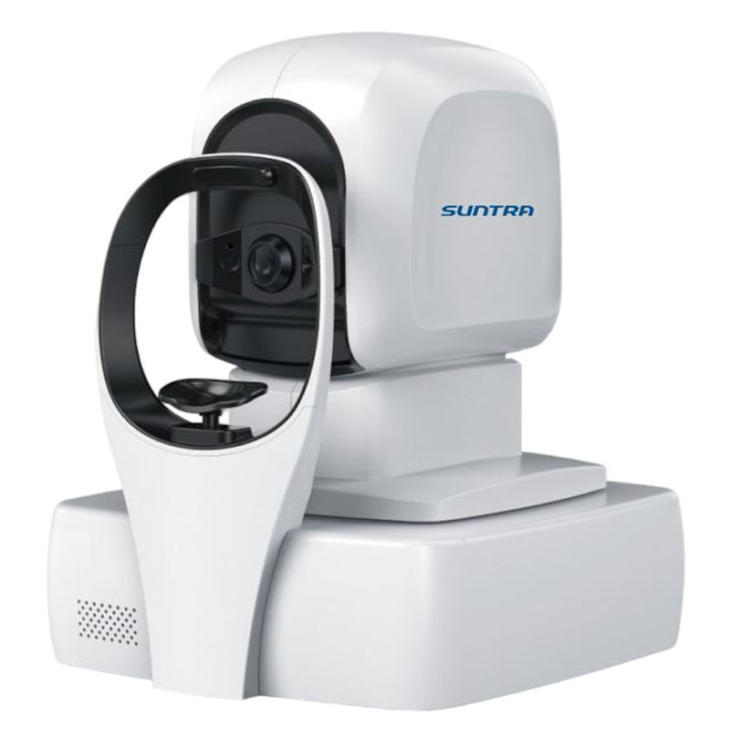
OCT-8200 Optical Coherence Tomography
OCT-8200 ophthalmic optical coherence tomography (OCT) is a fully automatic artificial intelligence OCT with fully independent intellectual property rights. Combined with multi-image registration and image enhancement technology and industry-leading analysis technology, it provides a full-stack solution for clinical diagnosis and treatment, including automatic image capture, accurate image analysis, intelligent eye health record management, and real-time data sharing on the cloud.
Technical Specification
| OCT-8200 | |
| Type | SD-OCT |
| Light energy (cornea) | ≤750μW |
| Measurement characteristic | Axial resolution:5μm Lateral resolution:20μm |
| Scanning characteristic | Maximum A-scan speed:≥20000times/second Scanning depth:2.3mm Maximum scanning range:12mm*9mm |
| Light Source characteristics | Central wavelength:840nm Optical power:≤750μW Refractive compensation range:-20D~+25D |
| Scanning mode | 5 kinds Large field of view: HD straight line, radiation six lines, local area: macula, optic disc |
| Scanning range | 12mm*9mm Complete the image acquisition of macula + optic disc at one time |
| Single line scan length | 12mm One scan macula + optic disc |
| Pupil requirement | 2mm |
| Automatic pupil orientation | √ |
| Automatic pupil compensation | √ |
| Fundus image | Full pixel imaging technology based on high scanning speed |
| Focusing mode | Fully automatic |
| Software analysis | Large field analysis Simultaneously covering macula and optic disc a total of 14 itemsLocal analysis – Optic disc 8 items Optic disc 6*6mm fundus image Cup-disc ratio Six lines Horizontal C/D Ratio Vertical C/D Ratio Optic cup area RNFL ring sweep thickness curve RNFL thickness topographic map TSNI thickness grid of nerve fiber layerLocal analysis – macular 8 items Macular 6*6mm fundus map Average thickness of the inner retina Average retinal thickness Macular fovea thickness ILM-RPE thickness topographic map ILM-RPE thickness grid RNFL thickness topographic map Retinal volume grid Anterior segment analysis (peripherals required) |
| User profile | √ |
| Integrated artificial intelligence | It can assist in the diagnosis of 16 major ophthalmic diseases, accurately identify, segment and automatically label the focal area |
| DICOM interface function | √ |
| Mode of operation | Fully automatic, manual operation |
Related Products
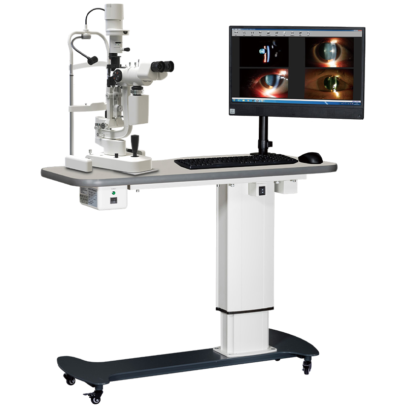
SMT-50T Slit Lamp Image System
The system is developed to meet the needs of ophthalmology users home and abroad,with a professional medical database;
It is used to analyze and process the photos and videos captured by the digital slit lamp,and to manage the information of the patients.
The microscope light path adopts the intersection Angle arallel design,good three-dimensional sense,wide field of vision;
Optical lens made of German optical glass,reduce optical distortion, claity and depth of field significantly improved;
All lenses are treated by multi-layer anti-reflection,moisture-proof and anti-mold coating,which always maintains good optical effect;
Imported light source,small heat, high brightness,uniform spot;
Background lighting system in dark room observation, shooting effect is especially obvious.
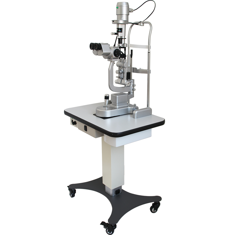
SMT-40D Slit Lamp Microscope
Galilean optical path provides high depth of view and large –angle observation
5-step drum magnifications, more convenient
All of the lens are multi-coated, moisture-proof and anti-reflection
Excellent optical performance gives clear image
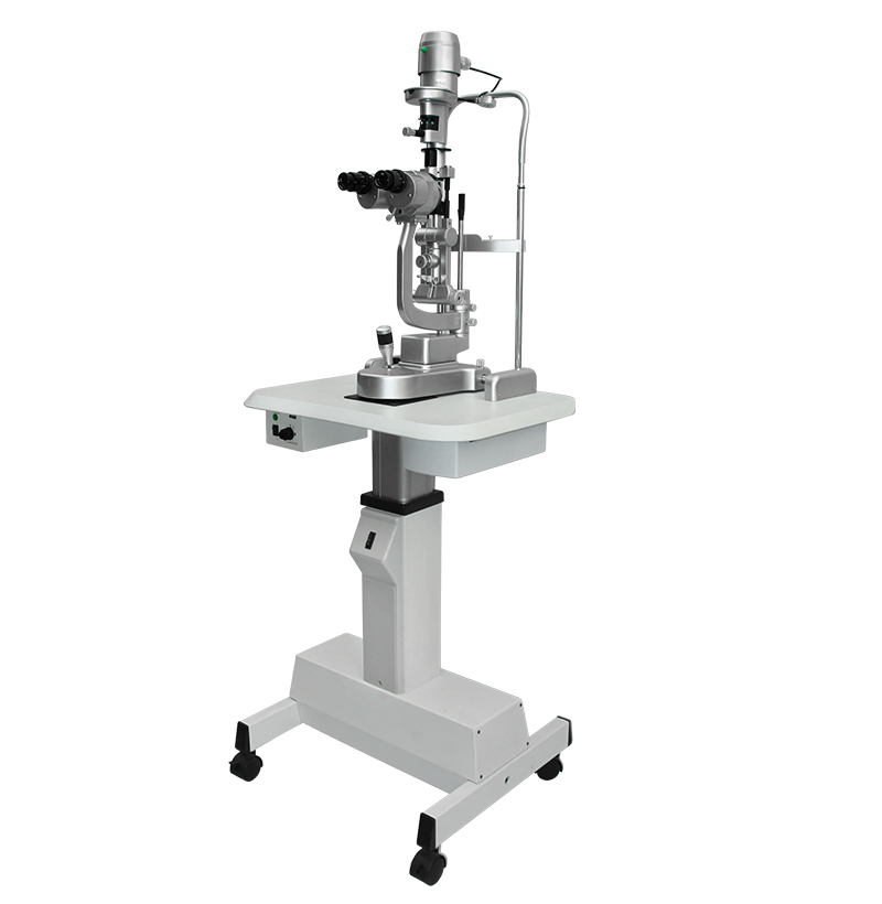
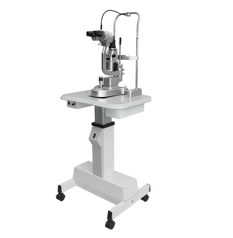
SMT-30S Slit Lamp Microscope
Universal slit lamp
3 step magnification: 10X, 16 X, 25X
Excellent optical performance gives a super clear optical image, afford broader field of view
14mm aperture
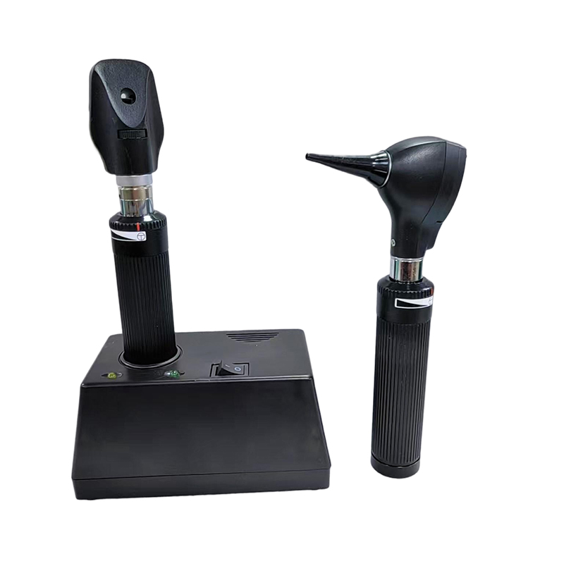
SMR-21&SMP-31 Rechargeable Ophthalmoscope & Otoscope Set
Share one desk charger and carry case;
Rechargeable Li-ion battery, Fully charged can be continuously used for more than 2 hours;
Adjustable brightness