ABOUT US
GET A FREE QUOTE
-
Home

-
Products
-
Medical Training Simulators
- Anatomy Education
Products
-
Medical Radiology

-
Ultrasound Scanner
-
Operating Room Equipment
-
Obstetric & Gynecology
-
Maternal & Infant Care
-
Sterilization Equipment
-
Diagnostic Equipment
-
ENT Equipment
-
Dental Equipment
-
Ophthalmic Equipment
-
In-Vitro Diagnostics
-
Hemodialysis Equipment
-
Laboratory Equipment
-
Medical Endoscopes
-
Hospital Furniture
-
Home Healthcare Equipment
-
First-Aid Equipment
-
Medical Training Simulators
-
Veterinary Equipment
-
Medical Refrigerator & Freezer
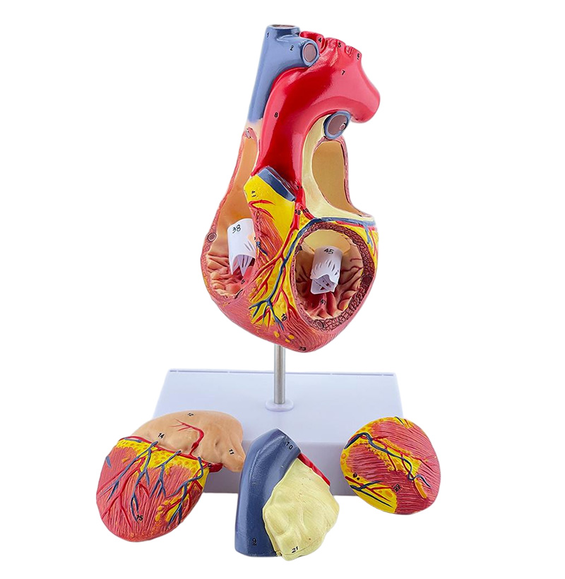
SYX04 Double the Heart
2-fold magnification heart model
The model consists of four parts including atrium and ventricle coronal section, right atrial appendage and left atrium, and displays the structures of atrium and ventricle as well as mitral valve, tricuspid valve, aortic valve and pulmonary valve, etc. The heart anatomical structure is elaborated in detail. A total of 59 site indicators are designed. On the basis of the original 1:1 model, the anatomical structure of atrial appendage is added. Besides, the left and right atria and ventricles can be disassembled to see the specific internal anatomical structure.
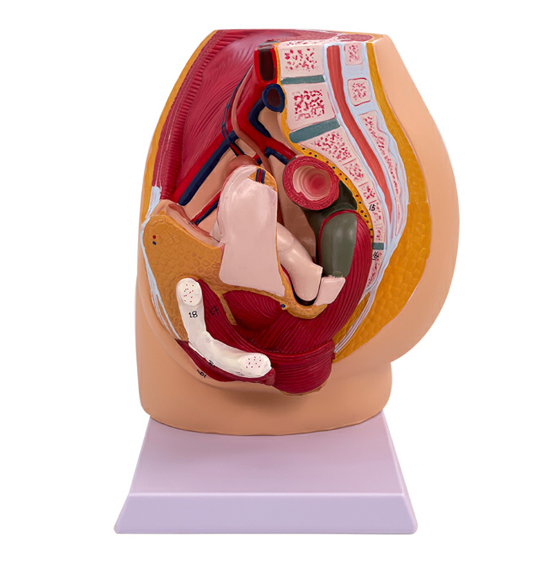
SYB32 Female Pelvic Sagittal
Female pelvic sagittal (4 parts)
This life-size model, showing all the important structures of the female pelvic system, and very detailed showing the abdominal cavity and pelvic muscles, the internal structure of the female genitourinary system is very detailed. All hand-painted and with digital indicators
The removable component includes a semi-open revealing medial and cross-sectional anatomy of the female reproductive system and uterus.
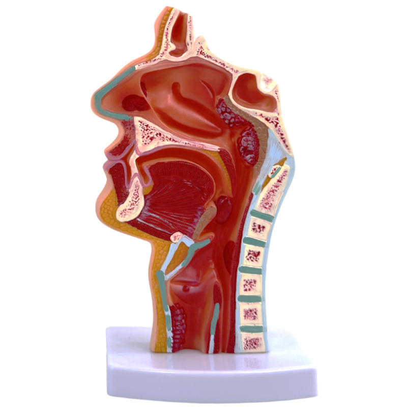
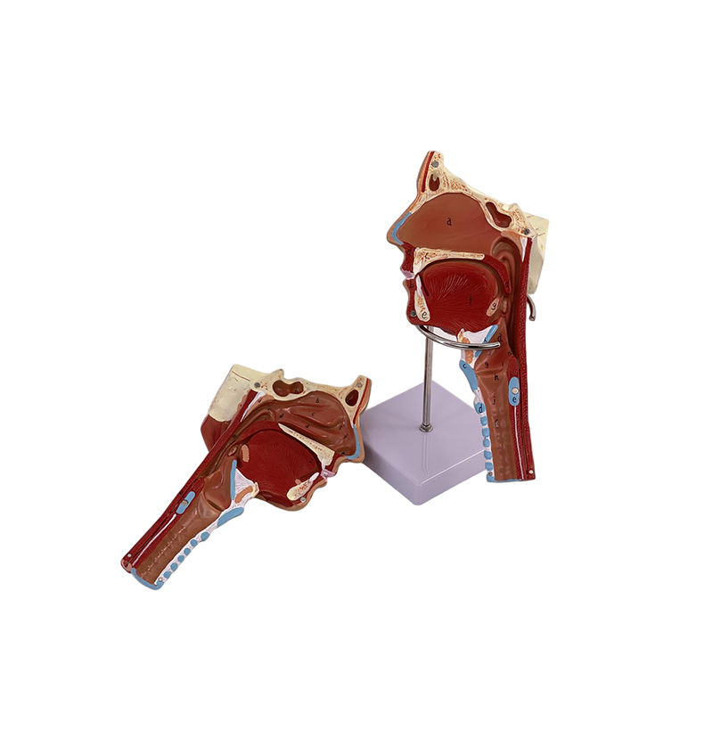
SY305C Pathological Model of Oropharynx
The pharynx of human body is divided into nasopharynx, oropharynx and hypopharynx. Nasopharynx is between skull base and soft palate plane.
And is communicated with the nasal cavity forward through the nasal posterior hole.
On both sides of nasopharynx, eustachian tube pharynx is communicated with middle ear and tympanum. Posterior pharyngeal recess is the most common site of nasopharyngeal ai.
The oropharynx is located between the soft palate and the superior margin of the epiglottis, and communicates with the oral cavity forward through the pharyngeal isthmus.
The out sidewall receives that palatine tonsil.
The hypopharynx is located between the upper margin of epiglottis and the plane of the lower margin of the 6th cervical vertebra, and communicates with the laryngeal cavity through the laryngeal orifice forwards and passes through the esophagus downwards.
The piriform recess on both sides of the laryngeal orifice is the place where the foreign body is easily retained.
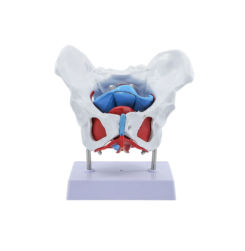
SY125 Female Pelvis And Pelvic Floor Muscle Model
Female pelvic and pelvic floor muscle model
This female pelvic and pelvic floor muscle model is suitable for medical schools to use as visual aids when explaining human anatomy, so that students can easily understand the muscle and fascia levels of the female pelvic region and perineum (pelvic diaphragm, urine reproductive diaphragm, etc.) and the location relationship of uterus, rectum and bladder.
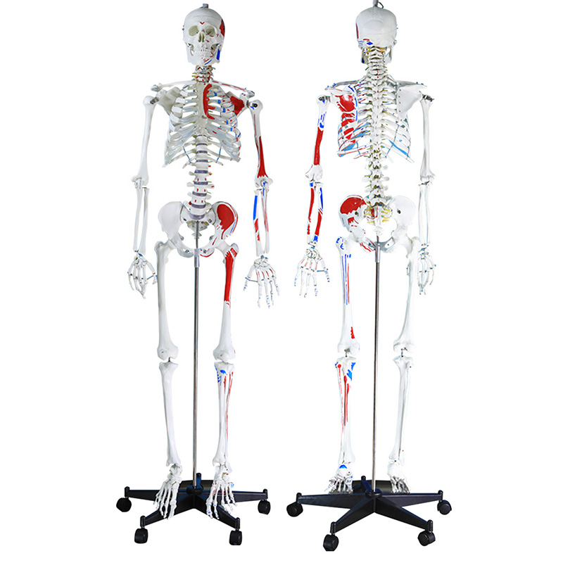
SY14 170 cm Frame with Muscle Coloration
170CM human skeletal model with half muscle staining and model
This economical, life-size, articulated human skeletal model is an ideal model for basic anatomy teaching at a good price. Arms and legs removable for study. Show nerve branches, spinal artery and lumbar intervertebral disc, etc. The whole model was stained with muscle attachment points on the half side of the human whole body skeletal model.
Material: PVC, imported paint. Iron base, stainless steel bracket.
Size: 170CM
J001 Pharyngeal Muscle Model
Environmental-friendly PVC material, with median cut for the design, using median cut design, bar model is divided into two parts, a detailed display of the human throat muscles and pharyngeal wall pharynx, submandibular gland and sublingual gland medial nasopharynx laryngeal anatomical structure, for understanding the throat muscles such as the cricopharyngeal muscle, nasopharynx cavity, glands and other structures of teaching and learning is a rare model.