ABOUT US
GET A FREE QUOTE
-
Home

-
Products
-
Medical Radiology
-
Digital X-ray Machine
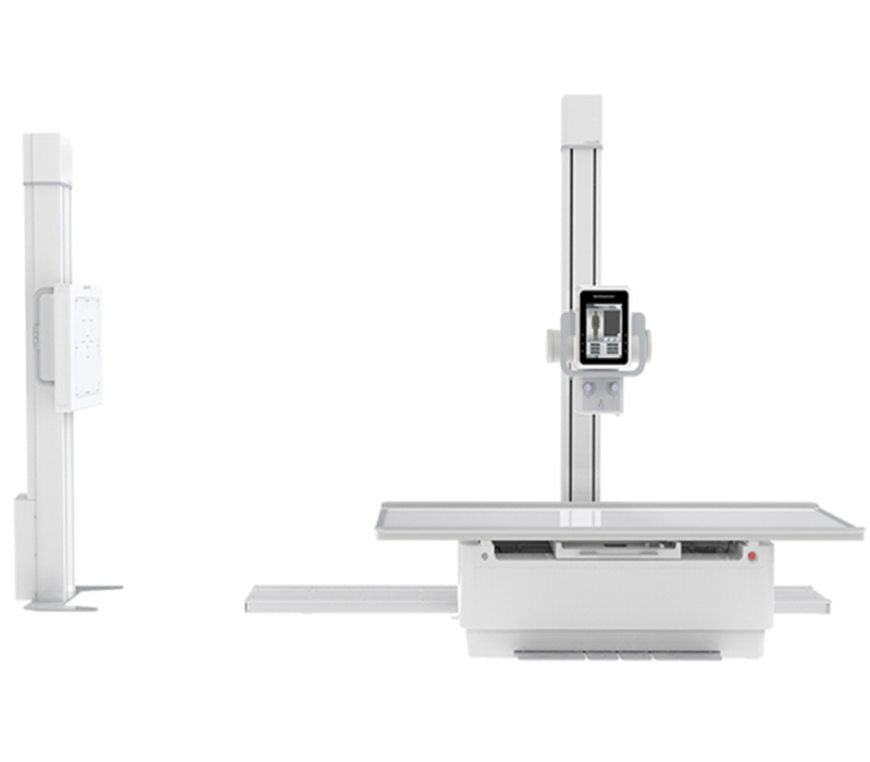
- SMT-050DE Double Column Digital Radiography System (Electric)
SMT-050DE Double Column Digital Radiography System (Electric)
Double column Digital Radiography System adopts large span independent ball tube frame design, easy installation, small occupation area, convenient maintenance. Four-way floating bed surface, easy to use and positioning. The lifting and lifting of X-ray source and bidirectional follow-up of horizontal position of bed column can reduce the positioning time and meet the requirements of large-flow clinical examination.
Applicable to orthopedics, emergency room, operating room, etc.,As human head, limbs, chest, spine, lumbar spine for medical diagnosis
Applicable to orthopedics, emergency room, operating room, etc.,As human head, limbs, chest, spine, lumbar spine for medical diagnosis
-
Features
1. Flexible mechanical system
Reasonable structural design, clear functional areas, and patient comfort
Humanized synchronous motion control, fast and accurate operation
2. Intelligent operation
The operation interface is highly integrated, and the image information platform is secure and reliable
Short exposure time, low radiation dose, providing safety assurance for patients and operators
3. High quality images
Strong penetration of radiation, high-quality images, and improved diagnostic efficiency for doctors
The system has image processing functions and management software to optimize the collected images
4. Efficient and Professional service team
24 hours service on-line
Technical Specification
Hardware Technical Specification
| Type | Item | Specification | |
| Generator | Output power | 50kW | |
| Inverter Frequency | 50kHz | ||
| kV | 40kV-150kV,1kV step | ||
| Current | 10-630mA | ||
| Exposure time | 1-6300ms | ||
| Exposure mAs | 0.1mAs~630mAs | ||
| X-ray tube | Type | LQ16-XD57-20.50 | Toshiba/E7843X |
| Focus | 0.6mm/1.2mm | 0.6mm/1.2mm | |
| Tube voltage | 40-150kV | 40-150kV | |
| Anode Heat capacity | ≥300kHU | ≥150kHU | |
| Tube Assembly
Heat Capacity |
≥1250kHU | ≥1250kHU | |
| Anode Speed | ≥2800rpm | ≥2700rpm | |
| Detector | Type | DT Imaging/PZ Medical/
Yu-imaging/iRay/CareRay |
|
| Material | CsI/A-Si | ||
| Imaging area | 17’’×17’’(43cm×43cm) | ||
| Pixel size | 139μm | ||
| Pixel matrix | 3072 × 3072 | ||
| Spatial resolution | 3.5 lp/mm | ||
| Image grey | 16bit | ||
| Image acquisition workstation | CPU | ≥3.0GHz,Windows system | |
| Hard disk | ≥1TB | ||
| Memory | ≥8GB | ||
| Display Card | ≥2GB | ||
| Monitor | 1600*1200Pixel Resolution,≥21〞 | ||
| Network interface | 100BaseT/1000BaseT,DICOM3.0 Transmission | ||
| Image storage capacity | ≥ 20,000 images | ||
| The pillar
with Tube |
Tube Vertical motion | Electric | |
| Tube Horizontal motion | Manual | ||
| Tube column horizontal rotation range | ±180° | ||
| Tube Rotation range | ±180° | ||
| Tube Longitudinal
Motion |
≥1500mm | ||
| Tube Lateral motion | ≥1900mm | ||
| Filtration | 1.0mmAL/70kv | ||
| SID | 1000mm-1800mm | ||
| Vertical photography stand
(chest X-ray stand) |
Lowest height detector center to ground | ≤470mm | |
| Detector center
motion range |
≥1550mm | ||
| Grid ratio | ≥10:1 | ||
| Grid density | ≥103LP/inch | ||
| Grid Focal length | ±180° | ||
| Tube Longitudinal
Motion |
≥180mm | ||
| Anti collision design | Yes | ||
| Patient table | Size | ≥2300mm x 800mm | |
| Load-bearing | ≥135KG | ||
| Permanent-magnet-locking control | |||
| 4-way floating | |||
| Power supply | 380VAC±10% 50/60Hz | ||
Software Technical Specification
| No. | Specification |
| I | Image acquisition and post-processing |
| 1 | Image preview function |
| 2 | Manual and automatic window width&level |
| 3 | Positive/negative film display |
| 4 | Reverse the horizontal and vertical positions of the image |
| 5 | Image rotation |
| 6 | Image movement and scaling |
| 7 | Image proportional cropping |
| 8 | Contrast enhancement |
| 9 | Image enlargement roaming |
| 10 | R/L annotation |
| 11 | Image storage |
| II | Patient management |
| 1 | Simple, advanced, or custom data query methods |
| 2 | Dicom standard image disc burning |
| 3 | Browse on any standard imaging workstation |
| 4 | Query and Management of Historical Image Data |
| 5 | Disk space detection, automatic cleaning of old inspection data |
| 6 | Dicom transmission of images seamlessly connected to hospital PACS |
| III | Software controllable exposure parameters |
| 1 | Manual/automatic/preset window width and level, local window width and level |
| 2 | Image positive and negative operations, image flipping, image rotation, image scaling, and roaming |
| 3 | Addition of Image information |
| 4 | Line, angle, and polygon measurement tools for images |
| 5 | Organizational balance, image contrast enhancement, pixel dose optimization |
| IV | Edge enhancement |
| 1 | Automatically recognize and analyze images |
| 2 | Enhance the sharpness of image edges |
| 3 | 1:1 preview of image post-processing |
| 4 | Enable doctors to make more intuitive diagnoses when optimizing images |
| V | Optimization of Dynamic range |
| 1 | Automatically compress the dynamic curves of the original image for easy diagnosis |
| 2 | High frequency image enhancement |
| 3 | Enhance image contrast, improve detail resolution, and significantly improve images such as bone trabeculae |
| VI | Noise suppression |
| 1 | Automatically filtering out unwanted signals, significantly reducing image noise, and improving image signal-to-noise ratio |
| VII | Film printing |
| 1 | Settings of Film properties, image layout, printing method |
| 2 | Quick typesetting of Manual/automatic |
| 3 | Choose any camera within the network |
| 4 | Customization of patient information and display location |
| 5 | Support queue management |
| 6 | Support settings of printing priority |
Package Information
| Item | Description | Length*width*height/mm | Weight/kg | Volume/m³ |
| Package1 | Patient table
Chest X-ray stand |
2270*1250*790 | 488 | 2.24 |
| Package2 | X-ray generator | 830*510*860 | 150 | 0.36 |
| Package3 | Computer,Detector
X-ray tube |
980*730*930 | 174 | 0.66 |
Standard Configuration
| No. | Configuration | Quantity |
| 1 | High frequency high voltage X-ray generator | 1 |
| 2 | X-ray tube | 1 |
| 3 | Flat panel detector | 1 |
| 4 | The pillar with Tube | 1 |
| 5 | Vertical photography stand (chest X-ray stand) | 1 |
| 6 | Beam limiter | 1 |
| 7 | Anti-Scatter Grid | 2 |
| 8 | Console | 1 |
| 9 | Image acquisition workstation | 1 |
| 10 | Power Cable | 1 |
| 11 | Patient table | 1 |
Related Products
.jpg)
SMT-3200MD Mobile Digital Radiography X-ray System
The machine is Combined X-ray photography of medical diagnostic equipment, application for wards, emergency rooms, operating rooms, ICU, etc. It can be used for human body head, limbs, chest, waist and abdomen and many other parts. It provides finest image resolution through Flat Panel detector. Easy operations make the work efficient.

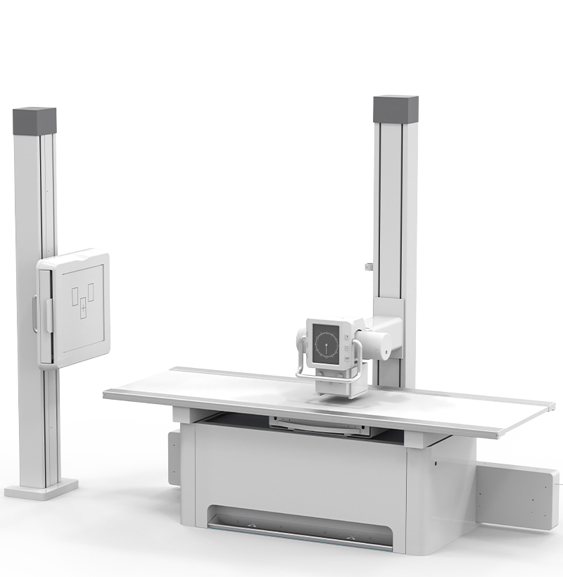
SMT-8000D Floor-mounted Digital Radiography System
80kW 1000mA Floor-mounted Digital X-ray Radiography System adopts floor-mounted table and wall stands. Its smart design, flexible movement, easy operation and high working efficiency provide more imaging positions. It is a high-end digital radiography device with wide range of applications, which could perform routine imaging of human head, chest, abdomen, lumbar vertebra and limbs with PA, LAT and clinostatism in hospitals at all levels.
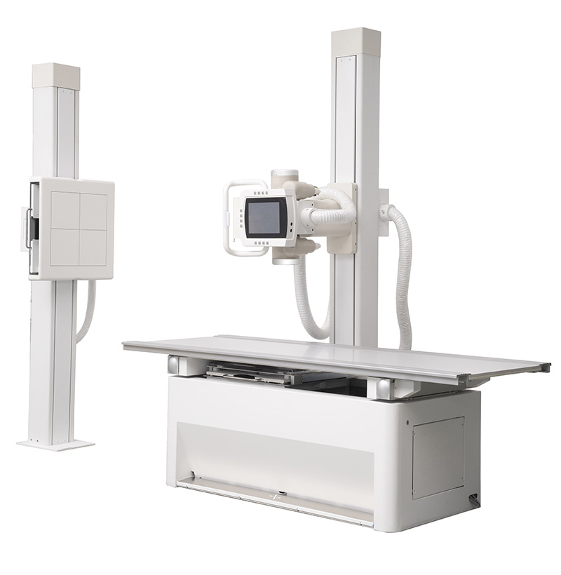
SMT-5000D Floor-mounted Digital Radiography System
50kW 630mA Floor-mounted Digital X-ray Radiography System adopts floor-mounted table and wall stands. Its smart design, flexible movement, easy operation and high working efficiency provide more imaging positions. It is a high-end digital radiography device with wide range of applications, which could perform routine imaging of human head, chest, abdomen, lumbar vertebra and limbs with PA, LAT and clinostatism in hospitals at all levels.

SMT-3200D Floor-mounted Digital Radiography System
Floor-mounted Digital X-ray Radiography System adopts floor-mounted table and wall stands. Its smart design, flexible movement, easy operation and high working efficiency provide more imaging positions. It is a high-end digital radiography device with wide range of applications, which could perform routine imaging of human head, chest, abdomen, lumbar vertebra and limbs with PA, LAT and clinostatism in hospitals at all levels.