ABOUT US
GET A FREE QUOTE
-
Home

-
Products
-
Medical Training Simulators
-
Emergency Skill Training
- J300 Electronic Human Tracheal Intubation Training Model
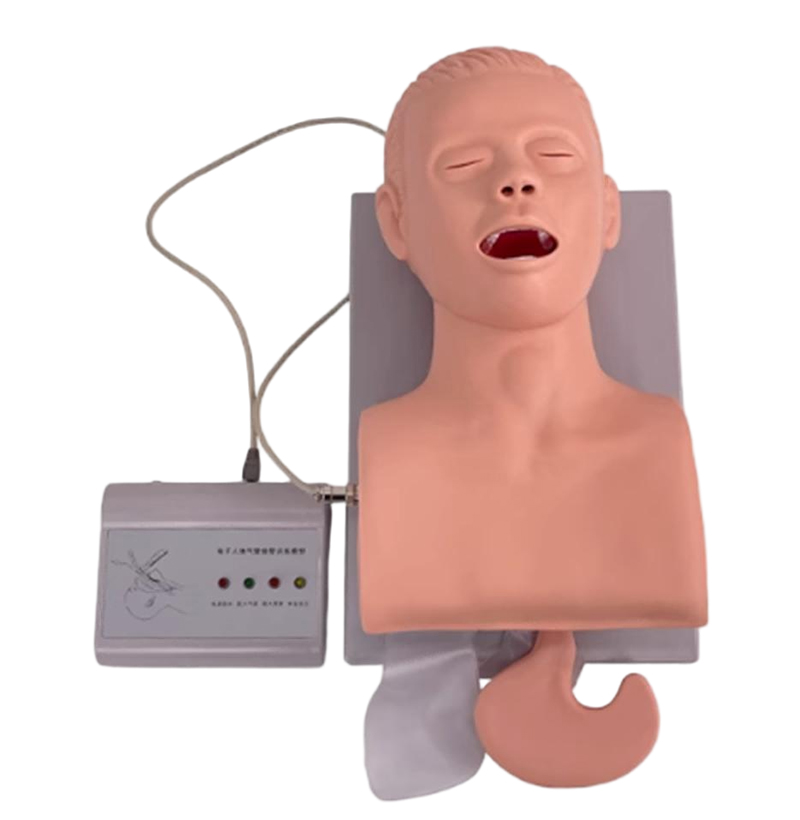
J300 Electronic Human Tracheal Intubation Training Model
It can be used for training operation and teaching demonstration of oral and nasal tracheal intubation.
During the training operation of oral and nasal tracheal intubation, the airway was inserted correctly, with electronic display and music playing functions; The air supply inflate both lungs and inject air into that tube. the balloon holds the tube.
During the training operation of oral and nasal tracheal intubation, the esophagus is inserted due to incorrect operation, with electronic display and alarm function. The gas supply distend that stomach.
During the training operation of oral and nasal tracheal intubation, the wrong operation causes the laryngoscope to compress teeth, with electronic display and alarm function.
Observe the pupils of normal pupils on one side and dilated pupils on the other side.
Indicates the location of the cricothyroid membrane puncture. Standard set configuration:
One set of adult standard head, neck and chest models
One electronic display
Features
It can be used for training operation and teaching demonstration of oral and nasal tracheal intubation.
■ During the training operation of oral and nasal tracheal intubation, the airway was inserted correctly, with electronic display and music playing functions; The air supply inflate both lungs and inject air into that tube. the balloon holds the tube.
■ During the training operation of oral and nasal tracheal intubation, the esophagus is inserted due to incorrect operation, with electronic display and alarm function. The gas supply distend that stomach.
■ During the training operation of oral and nasal tracheal intubation, the wrong operation causes the laryngoscope to compress teeth, with electronic display and alarm function.
■ Observe the pupils of normal pupils on one side and dilated pupils on the other side.
■ Indicates the location of the cricothyroid membrane puncture. Standard set configuration:
■ One set of adult standard head, neck and chest models
■ One electronic display
Related Products
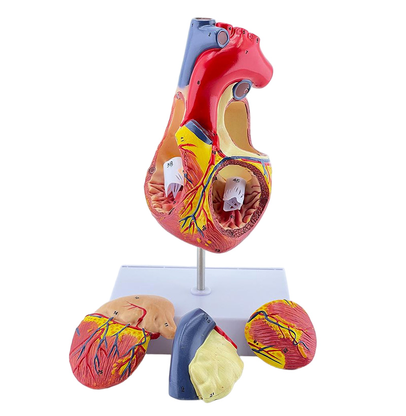
SYX04 Double the Heart
2-fold magnification heart model
The model consists of four parts including atrium and ventricle coronal section, right atrial appendage and left atrium, and displays the structures of atrium and ventricle as well as mitral valve, tricuspid valve, aortic valve and pulmonary valve, etc. The heart anatomical structure is elaborated in detail. A total of 59 site indicators are designed. On the basis of the original 1:1 model, the anatomical structure of atrial appendage is added. Besides, the left and right atria and ventricles can be disassembled to see the specific internal anatomical structure.
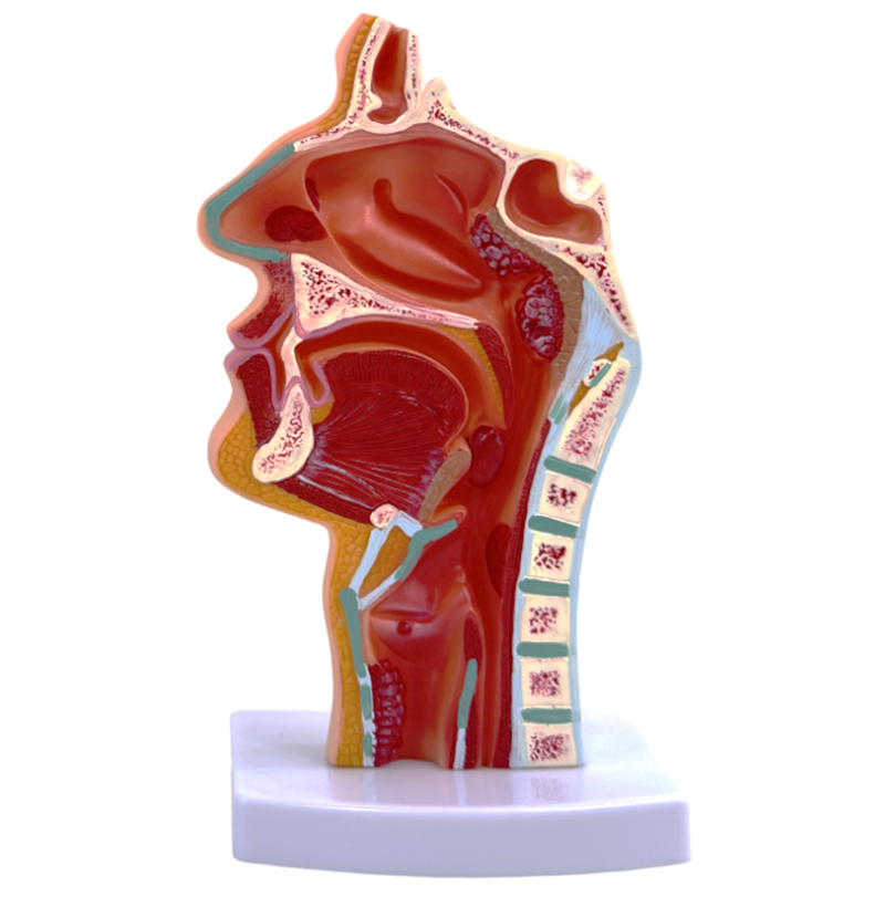
SY305C Pathological Model of Oropharynx
The pharynx of human body is divided into nasopharynx, oropharynx and hypopharynx. Nasopharynx is between skull base and soft palate plane.
And is communicated with the nasal cavity forward through the nasal posterior hole.
On both sides of nasopharynx, eustachian tube pharynx is communicated with middle ear and tympanum. Posterior pharyngeal recess is the most common site of nasopharyngeal ai.
The oropharynx is located between the soft palate and the superior margin of the epiglottis, and communicates with the oral cavity forward through the pharyngeal isthmus.
The out sidewall receives that palatine tonsil.
The hypopharynx is located between the upper margin of epiglottis and the plane of the lower margin of the 6th cervical vertebra, and communicates with the laryngeal cavity through the laryngeal orifice forwards and passes through the esophagus downwards.
The piriform recess on both sides of the laryngeal orifice is the place where the foreign body is easily retained.
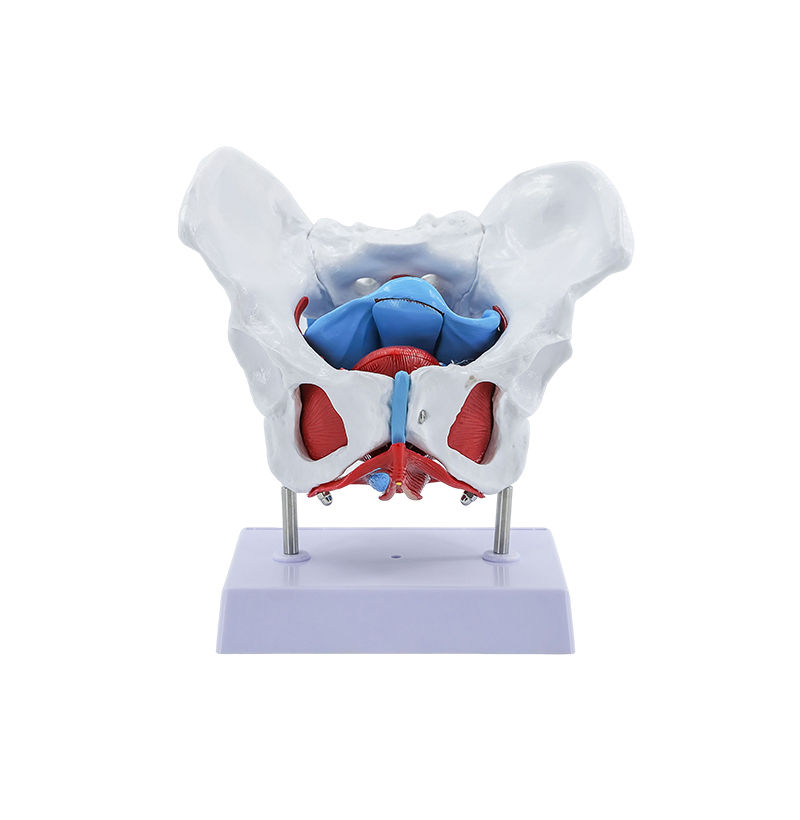
SY125 Female Pelvis And Pelvic Floor Muscle Model
Female pelvic and pelvic floor muscle model
This female pelvic and pelvic floor muscle model is suitable for medical schools to use as visual aids when explaining human anatomy, so that students can easily understand the muscle and fascia levels of the female pelvic region and perineum (pelvic diaphragm, urine reproductive diaphragm, etc.) and the location relationship of uterus, rectum and bladder.