ABOUT US
GET A FREE QUOTE
-
Home

-
Products
-
Ophthalmic Equipment
-
Ophthalmic Ultrasound
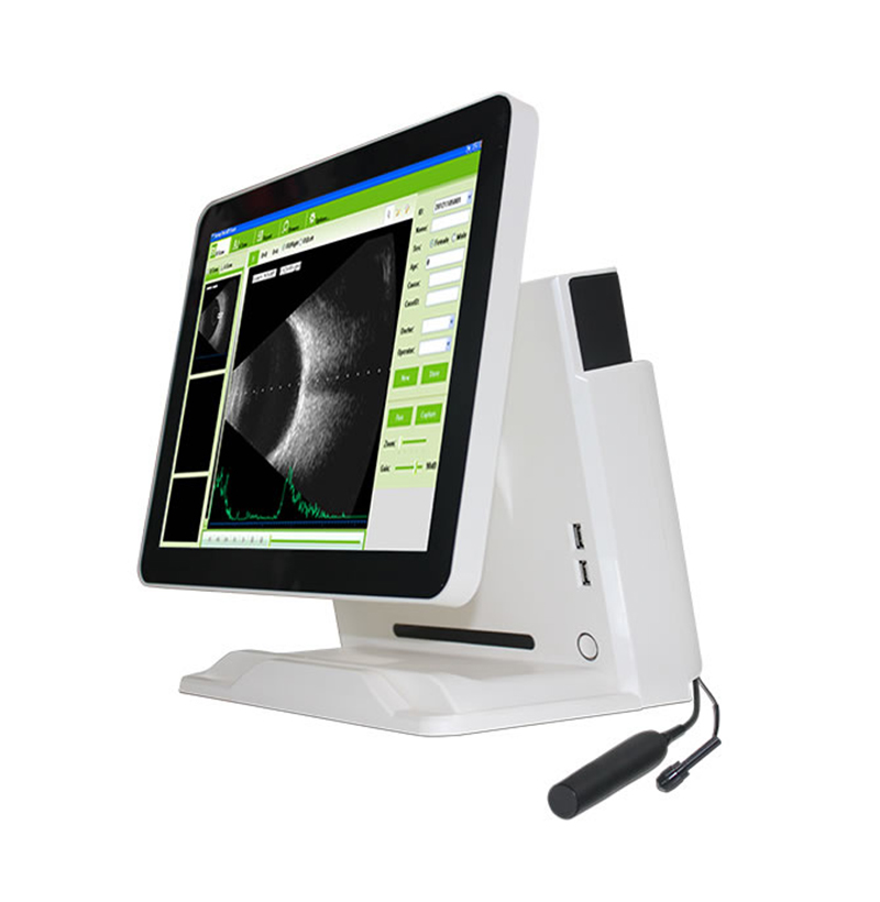
- SMT-2000U Ophthalmic A/B Scanner
Introduction
With normal, vitreous body enhancement, retina observation mode, mainly used for diagnosis of intraocular diseases, display the location, shape range of the focus of infection and the relationship with the surrounding tissue. Can be diagnosed vitreous opacity, retinal detachment, eye base tumors etc. eye diseases. A scan is used to measure anterior chamber depth, lens thickness, axial length, calculate diopter of implant IOL as well.
Technical Specification
| 1.General |
| Modules: |
| Ophthalmic Ultrasound B-Scan
Ophthalmic Biometry A-Scan |
| Features: |
| All in one device
15 inch LED touch screen Can work with Battery |
| Integrated Image
Capture Integrated Patient Database Integrated Report Editor |
| 2. A-Scan |
| Scan Modes: |
| Contact/Immersion |
| Examination Modes: |
| Normal Dense Cataract |
| Aphakic Pseudophakic(PMMA, Acrylic, Silicone) |
| Measurements: |
| AXL, ACD, Lens and Vitreous Individual Segment Velocities |
| Average and Standard Deviations for AXL, ACD, Lens&Vitreous |
| Specifications: |
| Clinical Accuracy ±0.1mm
Electrical Accuracy 0.0375mm |
| IOL Calculation in 0.5D Increments |
| IOL Calculation Formulas: |
| SRK-II SRK-T |
| Binkhorst-IⅡ Holladay |
| Hoffer-Q Haigis (Standard) |
| A-Scan Probe: |
| Hand-Held, Immersion or Slit Lamp Mounted Applicable |
| 3. B-Scan: |
| Scan Modes: |
| B Mode B+A Mode B+B Mode |
| Features: |
| Adjustable Zoom, Gain
Variable Gain Control |
| Capture of Frames and Cline Loops Available. |
| 256 Levels Gray Scale
Clinical Resolution: 0.1mm |
| Probe: |
| Transducer Frequency: 12.5MHz 53°Sector Scanning Method |
Related Products
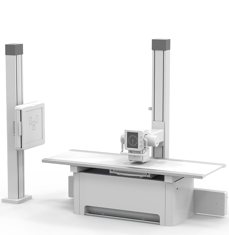
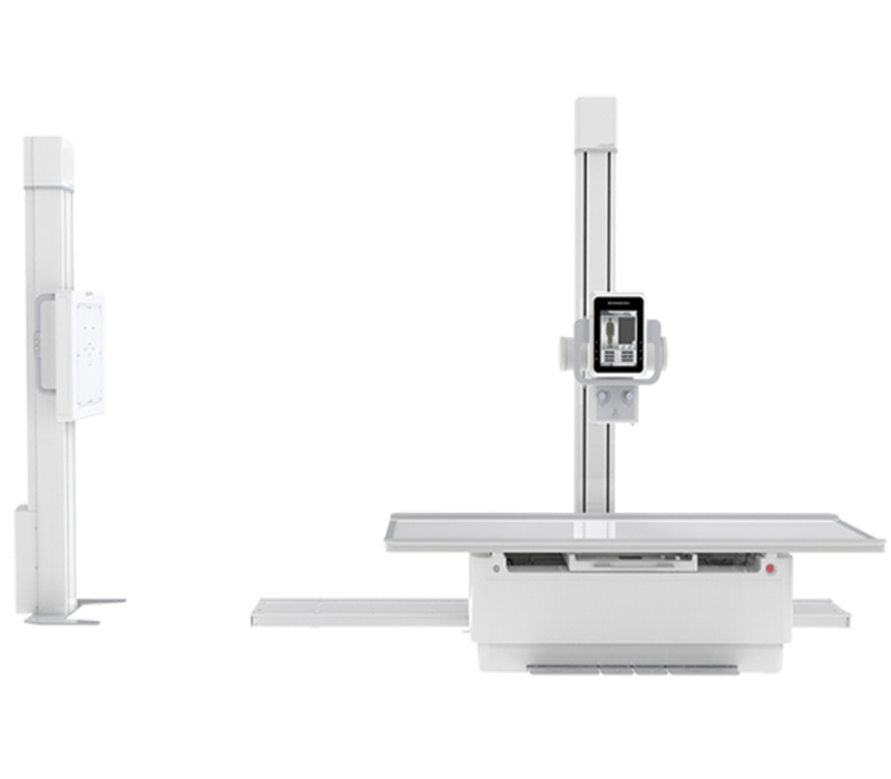
SMT-050DH Double column Digital Radiography System (Hand-driven)
Double column Digital Radiography System adopts large span independent ball tube frame design, easy installation, small occupation area, convenient maintenance. Four-way floating bed surface, easy to use and positioning. The lifting and lifting of X-ray source and bidirectional follow-up of horizontal position of bed column can reduce the positioning time and meet the requirements of large-flow clinical examination.
Applicable to orthopedics, emergency room, operating room, etc.,As human head, limbs, chest, spine, lumbar spine for medical diagnosis
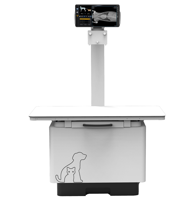
VSM-3200T 32kW 400mA Veterinary Digital Radiography System
1. Sophisticated technology, high rotating speed, good heat dissipation, high heat capacity, suitable for long-time clinical examination, long product life.
2. High frequency generator with high X-ray inverse frequency, large power and good X-ray quality.
3. Intelligence high voltage control system with various APR.
4. New generation a-Si Csl flat panel detector with high conversion rate achieves digital image acquisition with low dose.
5. High standard digital image processing system provides more post-processing and meets clinical needs.
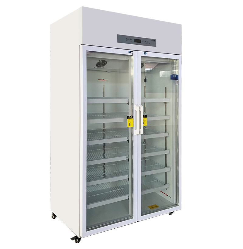
SM-800L 2-8°C Medicine Refrigerator
Meet the requirements of the new version GSP for medicine storage.
Can be used for medicine, reagents and various items that need cold storage.
Suitable for hospitals, clinics, blood stations of health and epidemic prevention systems, university laboratories, etc.
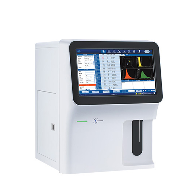
SMH-60B 5-part Auto Hematology Analyzer
1. Throughput 60T/H.
2. 14-inch touch screen.
3. Tri-angle laser scatter + flow cytometry method + impedance method for RBC and PLT counting.
4. 3D holographic scattergram displays the accurate 5-part differentiation of WBC.
5. Large storage capacity: 300,000 results(including histogram, scattergram, patient information).